Getting your Trinity Audio player ready...
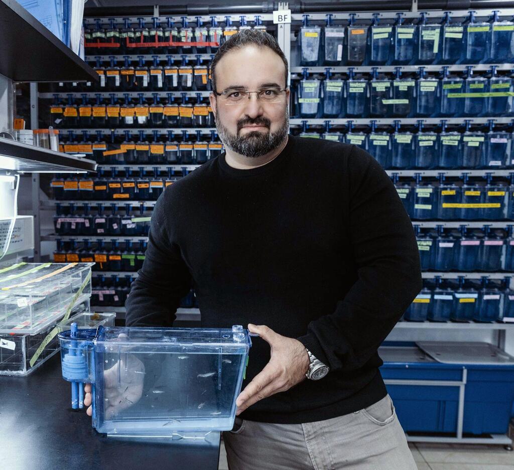
A new Israeli study reveals the fascinating mechanisms of egg cells as they prepare to create new life. The study, led by Dr. Yaniv Elkouby, a researcher in the Department of Developmental Biology at the Faculty of Medicine at the Hebrew University, and co-authors Swastik Kar and Rachael Deis from the Faculty of Medicine at the Hebrew University and the Institute for Medical Research, describes how developing eggs create cellular structures that organize the development of the embryo and provides important insights into processes essential to reproduction and developmental biology. The new findings were published in the Current Biology journal.
The key discovery: the Balbiani body
The focus of the study is the Balbiani body, a unique structure present in egg cells of a wide range of species, from insects to humans. The Balbiani body acts as an organizer of essential molecules, such as RNA and proteins, required for the proper development of the embryo.
In the new study, conducted using zebrafish models and cutting-edge imaging, the team discovered how this structure transforms from liquid droplets into a stable core, laying the groundwork for life itself. This discovery sheds light on the extraordinary precision of nature's reproductive process.
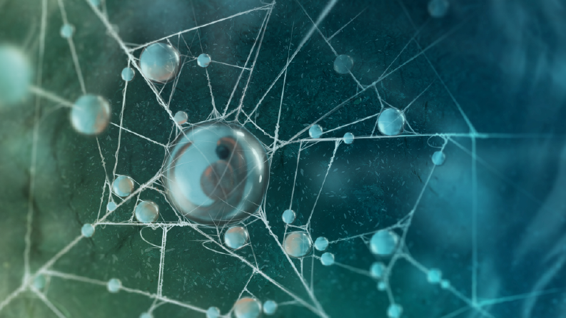
In this process, molecules made by the Balbiani body condense into viscous droplets that gradually merge to form a nearly solid organelle in the cell, a process that lays the foundations for the creation of life.
Prof. Elkouby explains: "This research focuses on understanding developmental processes - how a body develops with different cell types, structures and unique shapes that suit their function, from an egg that at first glance appears symmetrical, round and simple. This is an area that I also researched in my PhD, where I dealt with the development of the hindbrain in frogs. Every time I found an answer to a particular question, new questions emerged that led me deeper into the process of embryonic development.
"At a certain point, I realized that many of the factors that direct embryonic development originate in the egg. These processes are rooted in the egg already during its development in the ovary, and therefore my research focuses on understanding the mechanisms that shape these processes and lay the foundation for new life."
He adds that what particularly interested him was investigating the source of the information stored in the egg, and in his words: "Going as far back as possible during development to understand where this information comes from. This information, stored in the egg, is what directs the development of contractions in the embryo. At the right stage during development, this information comes into play and begins to shape the embryo's axes and body shape."
The researchers highlighted the role of a protein called "Bucky ball" that is created by the Balbiani body in a process called "molecular condensation." This process causes proteins to go from a soluble state in the cell to a solid-stable state, allowing the creation of a uniform and functional structure that separates its contents from the rest of the cell's environment. In the case of the Balbiani body, this separation "stores" the molecules of the Balbiani body's product for their later function in the embryo. This change is critical for the proper organization of the egg cell and the development of the embryo.
"Molecular condensation is a phenomenon that is being studied relatively recently in biological contexts, originating from studies in physics and chemistry," says Prof. Elkouby. "It was only in 2012 that studies began to be published that first revealed the existence of this phenomenon in cellular processes, and since then the field has developed to a wide range of biological connections.
To date, most studies in this field have been conducted on invertebrates, such as fruit flies and worms, or in cell cultures, and have made a tremendous contribution to its understanding. Our work is one of the first to demonstrate these mechanisms in a vertebrate organ during its development, a discovery that opens the door to a deep understanding of developmental processes. "
As for other proteins, Prof. Dr. Yaniv Elkouby work identified additional proteins that likely play important roles in the process of molecular condensation and organization of egg cells, but according to him, further research is needed to fully understand their roles and their impact on the process of embryonic development.
In addition, the study revealed the role of microtubules, cellular "cables" that regulate the movement of the protein Buckyball, form a scaffold for the fusion of its molecules and mechanically organize the correct shape of the Balbiani body. This process prevents errors in cell organization and ensures the success of the reproductive process.
Implications for reproductive health and disease
The study also offers a list of additional proteins, discovered through advanced proteomic approaches, that may play key roles in the process of Balbian formation. These proteins likely play important roles in the molecular condensation process and organization of egg cells, but according to Prof. Dr. Elkouby, further research is needed to fully understand their roles and their impact on the process of embryonic development. These findings pave the way for a deeper understanding of the mechanisms related to health, fertility and reproduction, especially in women.
In addition, the study also provides important insights into the mechanisms of molecular condensation in the cell and pathological processes. While solid structures in cells are often known in the context of degenerative diseases such as Alzheimer's and Parkinson's, the Balbian corpuscle is formed in a physiologically controlled manner, and decomposes when it completes its function. Understanding the control over the formation and decomposition of the corpuscle may lead to new insights into the mechanisms that cause neurodegenerative diseases.
According to Dr. Yaniv Elkouby: "We have been able to reveal how the Balbiani corpuscle is formed, organized and controlled at the molecular level. This discovery answers questions regarding the development of egg cells that have remained open for more than two centuries since the discovery of the corpuscle, and provides a new basis for research in the field of reproduction."
This discovery sheds new light on the processes of reproduction and embryonic development in vertebrates, including humans. The Balbiani corpuscle was first discovered in 1845 and is also formed in female eggs, but its functions and mechanisms of formation in female ovaries are still mysterious. The new findings provide tools for a better understanding of human reproduction and women's fertility and health.
Can the research findings be applied to understanding and improving fertility problems in women? "The Balbiani corpuscle is also formed in female eggs, and although studies in mice indicate its importance in follicle formation, the molecular understanding of its function is still incomplete," explains Prof. Elkouby.
"In female eggs, the structure of the corpuscle is very similar to that in zebrafish and mouse eggs, but the advantage of the zebrafish as a model system is particularly striking. Zebrafish serve as an excellent model for human biology, with a genetic similarity of about 70% and the ability to create accurate models of 80% of human genetic diseases, in a way that allows for research and finding a cure. Zebrafish ovaries are transparent and flat, which allows the use of high-resolution microscopy. Moreover, the zebrafish ovary contains almost all the critical stages of egg development, unlike mice where certain stages are not as accessible."
The findings of the study may help in the future to improve fertility problems in women, according to Prof. Elkouby: "In humans, the processes we are studying occur in the ovary of the developing embryo while it is still in its mother's womb. These stages are critical for determining the number and quality of eggs throughout a woman's life, since women are born with a fixed number of follicles.
Get the Ynetnews app on your smartphone: Google Play: https://bit.ly/4eJ37pE | Apple App Store: https://bit.ly/3ZL7iNv
"Defects in early egg development are a major cause of fertility problems, miscarriages and even the development of ovarian tumors, but currently there is a lack of basic understanding of the natural mechanisms that occur at this stage. Since it is impossible to study these processes directly in humans, we are using zebrafish to decipher these mechanisms at the most critical stages, thereby advancing our understanding of reproductive and developmental processes. Understanding the normal mechanisms will provide new insights into the causes of defects in these processes and ways to deal with them."


