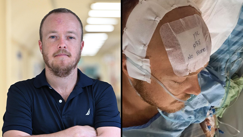
Three months after suffering a critical brain injury due to missile shrapnel, Ret. Capt. Saar Ram was operated on using 3D software that guided surgeons in replacing a shattered natural bone with a synthetic polymer implant to prevent further brain damage.
"I was called up on October 7 to the 53rd battalion of the 188th Brigade of the Armored corps. I arrived at the base in the north in the morning, and after two days we were moved to the envelope area, but it was not until the end of October that we entered the strip along with the Golani fighters," recounted Ram (29), who is studying industrial engineering and management. He engaged in combat from his tank against terrorists for three days before sustaining injuries.
"On November 3rd, a mortar shell or missile was fired at the tank, and from the blast, my head hit the metal and I broke my skull," he explains, "I remained conscious and didn't feel any pain in my head, only in my neck and shoulder, which we later diagnosed as dislocated. At the hospital, they explained to me that a frontal lobe injury does not produce pain because there are no nerve endings."
Ram acted swiftly, using his personal bandage to try and halt the bleeding by applying it tightly to his forehead. "But it didn't really help and the blood continued to flow," he shares, "since I was the only one injured in the tank, the crew moved me to the back, and my gunner took command and directed the tank to an evacuation APC that had a doctor. He treated me a bit in the field, an additional evacuation APC took me out of the strip, and from there a helicopter from Unit 669 took me to Rabin Medical Center." Despite the grave head injury and lack of sleep for three days prior to the incident, Ram managed to stay alert throughout the ordeal.
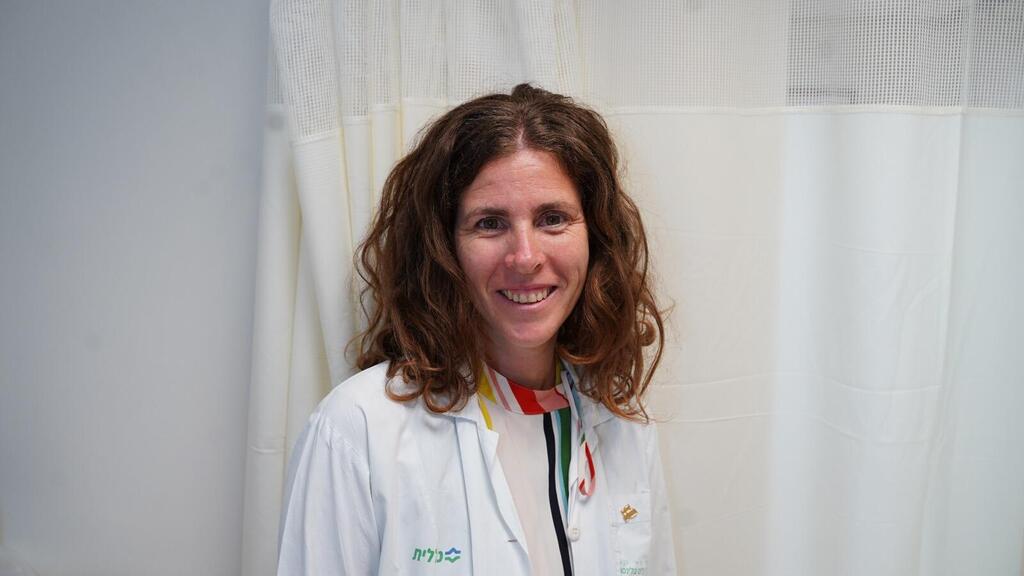
"Saar arrived with us severely injured," reports Dr. Idit Tamir, Director of the Functional Neurosurgery Service at the Department of Neurosurgery at Rabin Medical Center, "He had a severe head injury that resulted in a compressed open fracture, a condition that normally necessitates immediate surgery. However, because he was alert and the imaging showed no brain hemorrhage, we opted for conservative treatment initially: closing the skin defect on his forehead, allowing the area to heal, and monitoring his condition with further imaging."
A week later, once his life was deemed out of danger, he was allowed to go home for two weeks. "I was sleeping at my parents' house, and suddenly, in the middle of the night, a sharp pain in my shoulder woke me up," Ram recounts, "I took a painkiller, and when I got back to bed, I noticed it was a mess, with signs of urine and broken items next to it. I didn't understand what I was seeing, but I was alarmed and woke up my mother, and together we discovered that I also had bite marks on my tongue."
Ram was rushed to the hospital under suspicion of an epileptic seizure. "Epilepsy is a neurological disorder in the central nervous system that disrupts the brain's electrical activity, leading to excessive activity. It presents in the form of seizures and episodes that can range from mild and short-lived to severe, causing falls, loss of consciousness, and in extreme cases, can be fatal," Dr. Noa Shaikin, a neurologist specializing in epilepsy treatment at Assuta Medical Center, explains. "In some patients, the condition is genetic. In others, it can be triggered by a tumor, a cerebral event, infection, or head injury. Not all seizures mean the patient has epilepsy."
Dr. Tamir details, "He underwent a head CT, and we identified fluid of unknown origin at that point. It could have been either decomposed blood or an infectious fluid due to the open head wound. We proceeded to surgery, removed the bone fragments, sent a sample of the fluid for analysis, and closed the incision. The lab results indicated no infection, so he received medical treatment for potential seizures, was discharged home, and we scheduled a follow-up surgery using 3D technology."
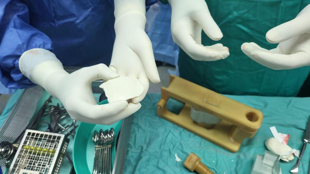
During the operation to remove the fluid and bone fragments, a CT scanner produced a three-dimensional image of his brain. "We forwarded this imaging outcome to a company that fashioned a precise mold of the missing bone area, reflecting his anatomical specifics," Dr. Tamir says.
On January 14th, Ram was back in surgery, emerging this time without a hole in his forehead and no longer at risk of brain exposure to infection due to the absence of the supporting bone. "While sedated and lying on his back, we opened the initial injury incision—from ear to ear, across the hairline. Picture an arch across the head, with incisions on each side," Dr. Tamir describes, "We then retracted the forehead skin from the virtual arch down to his eyes, and placed the polymer bone, secured to a titanium plate to avoid shifting, in the location of the original bone, thereby protecting the brain and sealing the forehead gap."
Dr. Tamir highlights the benefits of 3D technology in surgical procedures, noting it reduces operation times, minimizes risk of infection, and enhances the accuracy of outcomes. "Just a few years ago, surgeons handcrafted these implants," she notes, "It was akin to molding clay, and to ensure stability, we had to meticulously cut, sand, and adjust the implant. This process used to significantly extend the duration of surgery."
The integration of 3D software in medical practice has been accelerating since 2017, gradually leading to an increase in the printing of organs based on precise patient-specific anatomical models. Saar remains optimistic about his healing process. "Recovery from a brain injury is a gradual process," he mentions, "So the full extent of my injuries is still unclear. Although I face ongoing limitations and will always be considered disabled, I hope for the best."


