Getting your Trinity Audio player ready...
Mitochondria (singular: mitochondrion) are organelles within eukaryotic cells that generate most of the energy needed to power the various cellular processes and biochemical reactions. Unlike other organelles, mitochondria have their own genetic material, known as mitochondrial DNA (mtDNA), which is distinct from nuclear DNA—the primary genetic material of the organism, stored in the protected environment of the cell nucleus in eukaryotic organisms. Researchers hypothesize that in the distant past, mitochondria originated as free-living bacteria that entered into a symbiotic relationship with an ancestral eukaryotic cell and eventually became integral to it.
2 View gallery


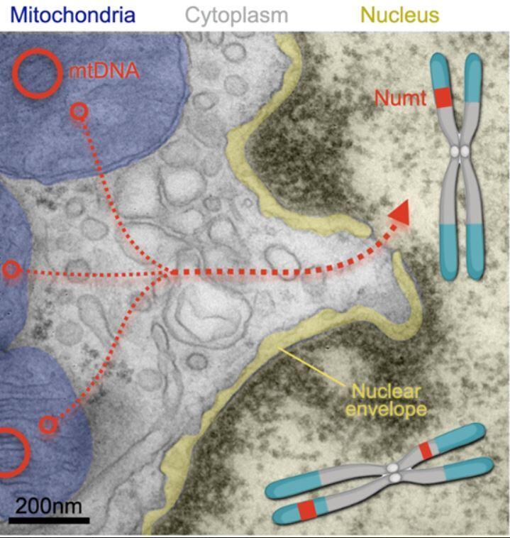
Segments of mitochondrial DNA have been found integrated into nuclear DNA in an increasing number of organisms. Illustration of mitochondrial DNA (mtDNA) entering the chromosome in the nucleus and forming NUMTs
(Photo: Martin Picard Laboratory, Columbia University Vagelos College of Physicians and Surgeons)
Over the past 30 years, it has become evident that the separation between mitochondrial DNA and nuclear DNA is not absolute. Segments of mitochondrial DNA have been discovered integrated into nuclear DNA across an growing number of organisms, ranging from unicellular fungi such as yeast to humans. This phenomenon is referred to as NUMT—an abbreviation for "nuclear mitochondrial DNA segment"—or mitochondria within the nucleus.
NUMTs have been studied very little in humans, and studies have primarily focused on white blood cells. The medical significance of NUMTs in cells remains unclear, though studies have reported a high accumulation of NUMTs in cancerous tumors. A new study has now expanded the research to the human brain, exploring potential links between NUMTs and neurodegenerative diseases.
Segments of mitochondrial DNA have been found integrated into nuclear DNA in an increasing number of organisms. Illustration of mitochondrial DNA (mtDNA) entering the chromosome in the nucleus and forming NUMTs | Image: Martin Picard Laboratory, Columbia University Vagelos College of Physicians and Surgeons
A look inside the brain
For the study, published in the journal PLOS Biology, researchers Ryan Mills from the University of Michigan and Martin Picard from Columbia University, analyzed genetic data from human brain cells. Samples were taken from the brains of individuals with neurodegenerative diseases and from those who had died from other causes, with their data preserved in genetic databases. In total, about 470 samples from the dorsolateral prefrontal cortex, 260 samples from the cerebellum, and approximately 70 samples from the occipital cortex were examined. Additionally, the researchers analyzed around 400 blood samples.
The researchers identified differences in NUMT accumulation across various tissues. Most cerebellum samples did not contain mitochondrial DNA in the cell nuclei, with an average of 0.75 NUMTs per sample. In contrast, occipital cortex samples averaged 1.7 NUMTs, increasing to 4.1 NUMTs per sample in the prefrontal cortex.In white blood cells, the average was 3.5 NUMTs per sample, with a much broader range: while NUMTs in brain samples did not exceed 30 per sample, some blood samples contained up to 300 NUMTs. Red blood cells, which lack nuclei, were not relevant to this study.
The researchers hypothesize that the differences between cerebellum and cortical samples are related to the number of mitochondria in each cell type. Cerebellar cells contain an average of 1,000 mitochondria per cell, while cells in the other two brain regions have about four times as many. However, the reason for the discrepancy between the prefrontal and occipital cortices remains unclear. In white blood cells, the number of mitochondria is lower—only a few hundred per cell—and the high NUMT count is likely due to their rapid division rate, whereas most neurons cease dividing at a young age.
The study did not find any correlation between the number of NUMTs in brain cells and neurodegenerative diseases. However, an unexpected finding was that healthy individuals who had died at a younger age had more NUMTs in the prefrontal cortex—on average, two additional NUMTs for every ten years of life lost. Currently, the researchers have no explanation for the correlation between the number of NUMTs and premature death.
Nuclear Mitochondria and aging
To explore the relationship between NUMTs and aging, researchers took cell samples from healthy individuals, cultured them, and monitored the integration of mitochondrial DNA into the nuclei over a period of up to seven months. Growing cells in culture mimics the process of aging to an extent: initially, the cells divide rapidly, approximately once every day and a half, but their division rate slows significantly over time, eventually occurring only about once a month.
The study found that the rate of NUMT integration into cell nuclei remained constant at an average of one NUMT every 12.6 days, regardless of the division rate, that is, the rate of aging. This rate increased slightly when the researchers added dexamethasone and oligomycin—drugs that affect mitochondrial activity. A much faster rate was observed when in cell cultures from patients with Leigh syndrome, a hereditary disorder involving severe mitochondrial dysfunction. In these cultures, the integration rate of mitochondrial DNA into cell nuclei rose to 3.7 NUMTs every 10 days.
Get the Ynetnews app on your smartphone: Google Play: https://bit.ly/4eJ37pE | Apple App Store: https://bit.ly/3ZL7iNv
The mechanism by which mitochondrial DNA fragments integrate into the protected genetic material of the cell nucleus remains unclear. However, the study, along with other research, suggests that mitochondrial damage increases the frequency of NUMT formation. This may occur because such damage releases DNA fragments from the mitochondria into the intracellular fluid, shortening their path to the nucleus. Once in the nucleus, the integration of these fragments into chromosomes may be facilitated by DNA repair mechanisms.
Much remains to be learned about the process of NUMTs formation (known as Nuntogenesis), their significance, and whether their accumulation in the brain contributes to early mortality or is merely a symptom of another underlying issue.


